Berlin Eyecare now offers Optomap retinal imaging!
When a five-year-old boy was blinded after a regular eye exam failed to spot a retinal detachment, his father made it his life’s work to help eye care professionals by revolutionizing retinal imaging. Optos was founded by Douglas Anderson with the goal to make a patient-friendly device to capture a digital ultra-widefield image of the retina. Today, millions of patients around the world have benefited from optomap® retinal imaging. You will find optomap systems in many independent optometrists, as well as hospitals, speciality clinics throughout the world and Harvard Medical Center in Massachusetts.
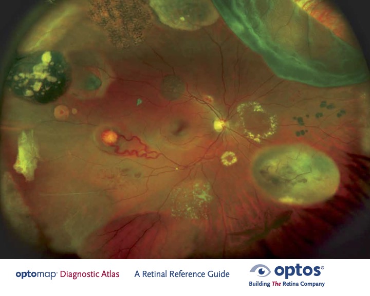
Optos has the capability to produce high resolution optomap images of 82% or 200◦ of the retina, something no other imaging device is capable of in a single comfortable capture. More than 2,500 published and ongoing clinical trials as well as thousands of case studies and testimonials show the long-term value of optomap imaging and OCT in diagnosis, treatment planning, and patient engagement.
What is an optomap Image?
Getting an optomap image is fast, painless, and comfortable. Nothing touches your eye at any time. It is suitable for the whole family. To have the exam, you simply look into the device one eye at a time (like looking through a keyhole) and you will see a flash of light to let you know the image of your retina has been taken.
Under normal circumstances, dilation drops might not be necessary, but your eye care practitioner will decide if your pupils need to be dilated depending on the health of your eyes.
Benefits of an optomap Image:
The benefits of having an optomap ultra-widefield retinal image taken are:
- optomap facilitates early protection from vision impairment or blindness
- personal comparison data to ensure no changes in your eye health from year to year
- Early detection of life-threatening diseases like cancer, stroke, and cardiovascular disease
The unique optomap ultra-widefield view helps your eye care practitioner detect early signs of retinal disease more effectively and efficiently than with traditional eye exams.
Early detection means successful treatments can be administered and reduces the risk to your sight and health.
Find out more about Optomap imaging and uses by visiting www.optos.com/patients/